- Main Page
- A1C Test
- Advance Directives
- Age on other Planets
- Aliens
- American Flag
- Annuals
- Anxiety
- Aortic Aneurysm
- Apple Cider Vinegar
- Arrhythmia
- Atrial Fibrillation
- Avoiding Scams
- Awareness Ribbons
- Bamboo
- Banana Tree, Grand Nain
- Banana Tree, Ice Cream
- Banana Tree, Zebrina Rojo
- Beekeeping
- Benign P P Vertigo
- Birth Month
- Blood Tests
- Blood Types
- Body Mass Index - BMI
- BMI Calculator
- Boogaloo
- Bookmarks
- Boot Anatomy
- Boot Fit Guide
- Boot Glossary
- Boot Leathers
- Boot Makers
- Boot Retailers
- Boot Styles - Western
- Boot Toes & Heels - Western
- Boot Toes & Heels - Work
- Bronchitis
- Candle Colors
- Carbohydrates
- Cardiac Catheterization
- Cardiovascular Disease
- CGM's
- Chakras
- Chinese Zodiac
- Cholesterol
- Christmas Tree
- Color Codes Chart
- C.O.P.D.
- Coronary Artery Disease
- Country Stars
- Cowboy Hat Etiquette
- Cowboy Hat Sizing
- C.P.A.P.
- Credit Score Checkers
- Crystals & Gems
- CT scan
- Degenerative Disk Disease
- Depression
- Diabetes Info.
- Diabetes Facts
- Diabetes - Pre
- Diabetes - Type 1
- Diabetes - Type 2
- Diabetes - Type 3c
- Diabetes - Gestational
- Diabetes Care
- Diabetes Care Team
- Diabetes Terms
- Diabetes Treatment
- Diabetes & Fruits
- Diabetes & Veg's
- Diet - Boiled Egg
- Diet - DASH
- Diet - Fat Burning
- Diet - Mediterranean
- Diet - Military
- Disability
- Do Not Resuscitate
- Dream Catchers
- Dupixent®
- Echocardiogram
- Electrocardiogram
- Emphysema
- Epsom Salt
- Eye Teasers
- Fairies
- Farxiga®
- Flower Astrology
- Fonts
- Foods To Regrow
- Friend
- Funny Things
- Fun Stuff
- Glycemic Index
- Gout
- Growing Blueberries
- Halloween
- Halloween Treats
- Headaches
- Health Info. Lines
- Heart Attack
- Heart Disease - Other
- Heart Failure
- Heart Tests
- Hello!!
- Herbal Codes
- Herbal Medicine
- Herb & Oils Uses
- Herniated disk
- Home Remedies
- House Plants
- Humalog®
- Hydrogen Peroxide
- Hyperglycemia
- Hypoglycemia
- Hyperkalemia
- Hypokalemia
- Hypertension
- Hypotension
- Important Numbers
- Insomnia
- Insulin
- Juice Recipes
- Karma
- Kidney Cysts
- Kidney Disease
- Kinds of Tea
- Lantus®
- Lemon Cleanse
- Logger vs Lineman
- Macaroni!!
- Medicare
- Mental Health
- MO HealthNet
- Moon Phases
- Mounjaro®
- MRI Scan
- My Athletic Shoes
- My Boots & Spurs
- My Cowboy Hats
- Myelography
- Mystical Unicorn
- Nasal Polyps
- Natal Astrology Chart
- Never Forget
- Nuclear Medicine
- Nutrition - Adults
- Nutrition - Adults, Older
- Nutrition - Kids
- Obesity
- One Little Rose
- Orchid Growing
- Orchid Sources
- Pagan Humor
- Pagans vs.Wiccans
- Parking Spaces
- PayPal.Me
- Pentagram vs. Pentacle
- Perennials
- Peripheral Artery Disease
- PET/CT Scan
- PET Scan
- Phobias A-Z
- Plant Care
- Plant Zone Map
- Potassium
- Propagating Plants
- Prurigo Nodularis
- Psychic Gifts
- PVC's
- Quit Smoking
- Recipes I like
- Red Yeast Rice
- Roses
- Runes
- Sadie & Beethoven
- Salt & Sodium
- Salt Water Flush
- Sciatica
- Service Animals
- Shape Shifters
- Sleep Apnea
- Sleep Disorders
- Sleep Studies
- Smile
- SPECT Scan
- Speed Test
- Spices You Need
- Spices I Have
- Spinal Stenosis
- Stents
- Steel Toe vs. Comp. Toe
- Stress Test - Exercise
- Stress Test - Nuclear
- Sugars - Sweeteners
- Superstitions
- Symbols
- Tarot
- The Ten Commandments
- Tools of the Craft
- Top Expensive Movies
- Top Modern Westerns
- Top 100 Westerns
- Toyota Yaris 2008
- Toyota Yaris 2012
- Trazodone
- Tree, Calamondin Orange
- Tree, Lemon (Meyer)
- Tree, Lime
- Tree Signs
- Ultrasound
- US Bill of Rights
- US Constitution
- US Declaration of Independence
- Vaccines by Age
- Vaccines 0-6 yrs
- Vaccines 7-18 yrs
- Vaccines 19 and up
- Ventricular Fibrillation
- Vertigo
- Vital Records
- Vital Signs
- Vitamin B12
- Vitamin C
- Vitamin D
- Vitamin E
- Vitamin K
- Vitamins & Minerals
- Water Therapy
- Weight on other Planets
- Wiccan Rede
- X-Rays
- Yin / Yang
- Zodiac Signs
Needed to read PDF's

X-Rays
Overview
An X-ray is a quick, painless test that captures images of the structures inside the body — particularly the bones.
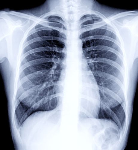
X-ray beams pass through the body. These beams are absorbed in different amounts depending on the density of the material they pass through. Dense materials, such as bone and metal, show up as white on X-rays. The air in the lungs shows up as black. Fat and muscle appear as shades of gray.
For some types of X-ray tests, a contrast medium — such as iodine or barium — is put into the body to get greater detail on the images.
What are X-rays?
X-rays use invisible electromagnetic energy beams to produce images of internal tissues, bones, and organs on film or digital media. Standard X-rays are performed for many reasons, including diagnosing tumors or bone injuries.
X-rays are made by using external radiation to produce images of the body, its organs, and other internal structures for diagnostic purposes. X-rays pass through body structures onto specially-treated plates (similar to camera film) or digital media and a "negative" type picture is made (the more solid a structure is, the whiter it appears on the film).
When the body undergoes X-rays, different parts of the body allow varying amounts of the X-ray beams to pass through. The soft tissues in the body (such as blood, skin, fat, and muscle) allow most of the X-ray to pass through and appear dark gray on the film or digital media. A bone or a tumor, which is more dense than soft tissue, allows few of the X-rays to pass through and appears white on the X-ray. When a break in a bone has occurred, the X-ray beam passes through the broken area and appears as a dark line in the white bone.
X-ray technology is used in other types of diagnostic procedures, such as arteriograms, computed tomography (CT) scans, and fluoroscopy.
Radiation during pregnancy may lead to birth defects. Always tell your radiologist or doctor if you suspect you may be pregnant.
Why it's done
X-ray technology is used to examine many parts of the body.
Bones and teeth
- Fractures and infections. In most cases, fractures and infections in bones and teeth show up clearly on X-rays.
- Arthritis. X-rays of the joints can show evidence of arthritis. X-rays taken over the years can help your healthcare team tell if your arthritis is worsening.
- Dental decay. Dentists use X-rays to check for cavities in the teeth.
- Osteoporosis. Special types of X-ray tests can measure bone density.
- Bone cancer. X-rays can reveal bone tumors.
Chest
- Lung infections or conditions. Evidence of pneumonia, tuberculosis or lung cancer can show up on chest X-rays.
- Breast cancer. Mammography is a special type of X-ray test used to examine breast tissue.
- Enlarged heart. This sign of congestive heart failure shows up clearly on X-rays.
- Blocked blood vessels. Injecting a contrast material that contains iodine can help highlight sections of the circulatory system so they can be seen easily on X-rays.
Abdomen
- Digestive tract issues. Barium, a contrast medium delivered in a drink or an enema, can help show problems in the digestive system.
- Swallowed items. If a child has swallowed something such as a key or a coin, an X-ray can show the location of that object.
How are X-rays performed?
X-rays can be performed on an outpatient basis, or as part of inpatient care.
Although each facility may have specific protocols in place, generally, an X-ray procedure follows this process:
The patient will be asked to remove any clothing or jewelry which might interfere with the exposure of the body area to be examined. The patient will be given a gown to wear if clothing must be removed.
The patient is positioned on an X-ray table that carefully positions the part of the body that is to be X-rayed--between the X-ray machine and a cassette containing the X-ray film or specialized image plate. Some examinations may be performed with the patient in a sitting or standing position.
Body parts not being imaged may be covered with a lead apron (shield) to avoid exposure to the X-rays.
The X-ray beam will be aimed at the area to be imaged.
The patient must be very still or the image will be blurred.
The technologist will step behind a protective window and the image is taken.
Depending on the body part under study, various X-rays may be taken at different angles, such as the front and side view during a chest X-ray.
Risks
Radiation exposure
Some people worry that X-rays aren't safe. This is because radiation exposure can cause cell changes that may lead to cancer. The amount of radiation you're exposed to during an X-ray depends on the tissue or organ being examined. Sensitivity to the radiation depends on your age, with children being more sensitive than adults.
Generally, however, radiation exposure from an X-ray is low, and the benefits from these tests far outweigh the risks.
However, if you are pregnant or suspect that you may be pregnant, tell your healthcare team before having an X-ray. Though most diagnostic X-rays pose only small risk to an unborn baby, your care team may decide to use another imaging test, such as ultrasound.
Contrast medium
In some people, the injection of a contrast medium can cause side effects such as:
- A feeling of warmth or flushing.
- A metallic taste.
- Lightheadedness.
- Nausea.
- Itching.
- Hives.
Rarely, severe reactions to a contrast medium occur, including:
- Very low blood pressure.
- Difficulty breathing.
- Swelling of the throat or other parts of the body.
How you prepare
Different types of X-rays require different preparations. Ask your healthcare team to provide you with specific instructions.
What to wear
In general, you undress whatever part of your body needs examination. You may wear a gown during the exam depending on which area is being X-rayed. You also may be asked to remove jewelry, eyeglasses and any metal objects because they can show up on an X-ray.
Contrast material
Before having some types of X-rays, you're given a liquid called contrast medium. Contrast mediums, such as barium and iodine, help outline a specific area of your body on the X-ray image. You may swallow the contrast medium or receive it as an injection or an enema.
What you can expect
During the X-ray
X-rays are performed at medical offices, dentists' offices, emergency rooms and hospitals — wherever an X-ray machine is available. The machine produces a safe level of radiation that passes through the body and records an image on a specialized plate. You can't feel an X-ray.
A technologist positions your body to get the necessary views. Pillows or sandbags may be used to help you hold the position. During the X-ray exposure, you remain still and sometimes hold your breath to avoid moving so that the image doesn't blur.
An X-ray procedure may take just a few minutes for a simple X-ray or longer for more-involved procedures, such as those using a contrast medium.
Your child's X-ray
If a young child is having an X-ray, restraints or other tools may be used to keep the child still. These won't harm the child and they prevent the need for a repeat procedure, which may be necessary if the child moves during the X-ray exposure.
You may be allowed to remain with your child during the test. If you remain in the room during the X-ray exposure, you'll likely be asked to wear a lead apron to shield you from unnecessary X-ray exposure.
After the X-ray
After an X-ray, you generally can resume usual activities. Routine X-rays usually have no side effects. However, if you're given contrast medium before your X-ray, drink plenty of fluids to help rid your body of the contrast. Call your healthcare team if you have pain, swelling or redness at the injection site. Ask your team about other symptoms to watch for.
Results
X-rays are saved digitally on computers and can be viewed on-screen within minutes. A radiologist typically views and interprets the results and sends a report to a member of your healthcare team, who then explains the results to you. In an emergency, your X-ray results can be made available in minutes.
One Final Note..
X-rays are one of the oldest, most reliable medical technologies. And despite it being almost 130 years since their discovery, they’re still relevant thanks to research into new, better ways to use them. Modern X-rays are far more detailed and use less radiation than in the past, thanks to advances in imaging resolution.
X-rays allow providers to quickly check what might be going on inside your body. This means you can know fast whether there’s something concerning that needs treatment or additional testing. Let your provider know if you have any questions about getting an X-ray or the findings.
Find me on Social Media


 |
Don't forget to bookmark me to see updates.. Copyright © 2000 - 2025 K.
Kerr |