- Main Page
- A1C Test
- Advance Directives
- Age on other Planets
- Aliens
- American Flag
- Annuals
- Anxiety
- Aortic Aneurysm
- Apple Cider Vinegar
- Arrhythmia
- Atrial Fibrillation
- Avoiding Scams
- Awareness Ribbons
- Bamboo
- Banana Tree, Grand Nain
- Banana Tree, Ice Cream
- Banana Tree, Zebrina Rojo
- Beekeeping
- Benign P P Vertigo
- Birth Month
- Blood Tests
- Blood Types
- Body Mass Index - BMI
- BMI Calculator
- Boogaloo
- Bookmarks
- Boot Anatomy
- Boot Fit Guide
- Boot Glossary
- Boot Leathers
- Boot Makers
- Boot Retailers
- Boot Styles - Western
- Boot Toes & Heels - Western
- Boot Toes & Heels - Work
- Bronchitis
- Candle Colors
- Carbohydrates
- Cardiac Catheterization
- Cardiovascular Disease
- CGM's
- Chakras
- Chinese Zodiac
- Cholesterol
- Christmas Tree
- Color Codes Chart
- C.O.P.D.
- Coronary Artery Disease
- Country Stars
- Cowboy Hat Etiquette
- Cowboy Hat Sizing
- C.P.A.P.
- Credit Score Checkers
- Crystals & Gems
- CT scan
- Degenerative Disk Disease
- Depression
- Diabetes Info.
- Diabetes Facts
- Diabetes - Pre
- Diabetes - Type 1
- Diabetes - Type 2
- Diabetes - Type 3c
- Diabetes - Gestational
- Diabetes Care
- Diabetes Care Team
- Diabetes Terms
- Diabetes Treatment
- Diabetes & Fruits
- Diabetes & Veg's
- Diet - Boiled Egg
- Diet - DASH
- Diet - Fat Burning
- Diet - Mediterranean
- Diet - Military
- Disability
- Do Not Resuscitate
- Dream Catchers
- Dupixent®
- Echocardiogram
- Electrocardiogram
- Emphysema
- Epsom Salt
- Eye Teasers
- Fairies
- Farxiga®
- Flower Astrology
- Fonts
- Foods To Regrow
- Friend
- Funny Things
- Fun Stuff
- Glycemic Index
- Gout
- Growing Blueberries
- Halloween
- Halloween Treats
- Headaches
- Health Info. Lines
- Heart Attack
- Heart Disease - Other
- Heart Failure
- Heart Tests
- Hello!!
- Herbal Codes
- Herbal Medicine
- Herb & Oils Uses
- Herniated disk
- Home Remedies
- House Plants
- Humalog®
- Hydrogen Peroxide
- Hyperglycemia
- Hypoglycemia
- Hyperkalemia
- Hypokalemia
- Hypertension
- Hypotension
- Important Numbers
- Insomnia
- Insulin
- Juice Recipes
- Karma
- Kidney Cysts
- Kidney Disease
- Kinds of Tea
- Lantus®
- Lemon Cleanse
- Logger vs Lineman
- Macaroni!!
- Medicare
- Mental Health
- MO HealthNet
- Moon Phases
- Mounjaro®
- MRI Scan
- My Athletic Shoes
- My Boots & Spurs
- My Cowboy Hats
- Myelography
- Mystical Unicorn
- Nasal Polyps
- Natal Astrology Chart
- Never Forget
- Nuclear Medicine
- Nutrition - Adults
- Nutrition - Adults, Older
- Nutrition - Kids
- Obesity
- One Little Rose
- Orchid Growing
- Orchid Sources
- Pagan Humor
- Pagans vs.Wiccans
- Parking Spaces
- PayPal.Me
- Pentagram vs. Pentacle
- Perennials
- Peripheral Artery Disease
- PET/CT Scan
- PET Scan
- Phobias A-Z
- Plant Care
- Plant Zone Map
- Potassium
- Propagating Plants
- Prurigo Nodularis
- Psychic Gifts
- PVC's
- Quit Smoking
- Recipes I like
- Red Yeast Rice
- Roses
- Runes
- Sadie & Beethoven
- Salt & Sodium
- Salt Water Flush
- Sciatica
- Service Animals
- Shape Shifters
- Sleep Apnea
- Sleep Disorders
- Sleep Studies
- Smile
- SPECT Scan
- Speed Test
- Spices You Need
- Spices I Have
- Spinal Stenosis
- Stents
- Steel Toe vs. Comp. Toe
- Stress Test - Exercise
- Stress Test - Nuclear
- Sugars - Sweeteners
- Superstitions
- Symbols
- Tarot
- The Ten Commandments
- Tools of the Craft
- Top Expensive Movies
- Top Modern Westerns
- Top 100 Westerns
- Toyota Yaris 2008
- Toyota Yaris 2012
- Trazodone
- Tree, Calamondin Orange
- Tree, Lemon (Meyer)
- Tree, Lime
- Tree Signs
- Ultrasound
- US Bill of Rights
- US Constitution
- US Declaration of Independence
- Vaccines by Age
- Vaccines 0-6 yrs
- Vaccines 7-18 yrs
- Vaccines 19 and up
- Ventricular Fibrillation
- Vertigo
- Vital Records
- Vital Signs
- Vitamin B12
- Vitamin C
- Vitamin D
- Vitamin E
- Vitamin K
- Vitamins & Minerals
- Water Therapy
- Weight on other Planets
- Wiccan Rede
- X-Rays
- Yin / Yang
- Zodiac Signs
Needed to read PDF's

SPECT Scan
Overview
A Single-photon emission computed tomography (SPECT) scan is a specialized imaging test that produces detailed, three-dimensional images of blood flow to tissues and organs. This advanced diagnostic technique is a key component of nuclear medicine, which uses small amounts of radioactive substances to evaluate organ function and detect abnormalities. SPECT imaging is widely utilized to diagnose and manage conditions affecting the brain, heart, and other vital organs.
First introduced in the 1960s, SPECT scans have evolved significantly over the decades, becoming a cornerstone of modern medical imaging. By integrating nuclear medicine with computed tomography (CT), SPECT scans offer unique insights into both the structure and function of tissues, making them invaluable for identifying diseases at an early stage.
What is a SPECT scan?
Single-photon emission computerized tomography (SPECT) scans use radioactive materials and a specially designed camera to produce three-dimensional images of your organs and tissues.
This type of imaging provides a non-invasive way for healthcare providers to evaluate the health of certain parts of your body, most commonly the heart, brain, and bones.
What makes SPECT scans different from other methods of imaging is that it can show how well certain organs are functioning. For example, the images made by the SPECT scan can help pinpoint the location of seizures in people with epilepsy and assess whether there's sufficient blood flow to different areas of the brain.
Read on to learn why your healthcare provider may order this, who shouldn't have a SPECT scan, what to expect if you get one done, and how the results are interpreted.
What does a SPECT scan show?
In addition to showing what the structures inside your body look like, a SPECT scan shows how blood flows through your arteries and veins to your organs and tissues.
The most common types of SPECT scans include:
- SPECT brain scan. This helps assess and diagnose neurological disorders such as epilepsy, dementia, Parkinson’s disease, strokes and brain injuries. It can also show which brain areas have more or less activity and how much blood is flowing to them.
- SPECT heart scan. Conditions that may be revealed with a heart scan include ineffective circulation, scar tissue on the heart (such as from a heart attack), clogged or narrowed arteries and other heart abnormalities. It can also assess how well a patient is healing from heart surgeries and procedures.
- SPECT bone scan. Conditions that may be revealed with a bone scan include bone infections, cancer that has spread to the bones or bone fractures.
Depending on your situation, your provider may do a regular SPECT scan or a SPECT/CT scan. A SPECT/CT scan combines two types of scans (single photon emission computed tomography and computed tomography).
SPECT brain scan
A SPECT test creates a detailed, 3D map of the blood flow activity in the brain, It can tell your provider which areas of your brain are most active and least active.
SPECT brain scans help diagnose neurological conditions like:
- Seizure disorders. A SPECT scan can help diagnose and treat seizure disorders, such as epilepsy. It does this by pinpointing the area of seizure activity in the brain
- Clogged blood vessels. SPECT scanning can find issues with blood flow in the brain. It can help diagnose or check on vascular brain disorders, such as moyamoya disease, a condition in which the arteries in the brain become blocked or narrowed.
- Parkinson’s disease. Parkinson’s disease is a movement disorder caused by the degeneration of dopamine-producing neurons in the brain. Symptoms include tremors, muscle rigidity, and slowed movement. SPECT imaging, particularly dopamine transporter (DaT) scans, detects abnormalities in dopamine activity, aiding in the diagnosis of Parkinson’s disease. This test is especially valuable for differentiating Parkinson’s from other movement disorders. With a PPV of around 88% and an NPV of 92%, SPECT scans provide critical insights into disease progression and help guide treatment decisions.
- Epilepsy. Epilepsy is a neurological condition characterized by recurrent seizures. Identifying the specific brain regions responsible for seizure activity is crucial for effective treatment planning, particularly for patients considering surgery. A SPECT scan for epilepsy employs a technique called ictal SPECT, which captures images during or immediately after a seizure. This approach helps pinpoint areas of abnormal blood flow in the brain. SPECT imaging has a PPV of 80-90% and an NPV of 85% in localizing seizure foci, making it an indispensable tool for pre-surgical evaluation.
- Alzheimer’s disease. Alzheimer’s disease is a progressive neurological disorder that leads to memory loss, cognitive decline, and behavioral changes. Early and accurate diagnosis is essential for managing symptoms and planning care. A SPECT scan for Alzheimer’s diagnosis detects areas of reduced blood flow in specific brain regions, such as the parietal and temporal lobes, which are commonly affected by the disease. Research indicates that SPECT imaging has a PPV of approximately 85% and an NPV of 90% for Alzheimer’s, making it a reliable tool for distinguishing Alzheimer’s from other forms of dementia.
- Traumatic brain injuries (TBIs).
- Dementia
SPECT heart scan
Because the radioactive tracer highlights areas of blood flow, this type of SPECT scan can show your provider how well your heart works. It can help diagnose a wide range of conditions, including:
- Scar tissue, which can indicate a heart attack.
- Coronary artery disease (narrowed arteries).
- Atherosclerosis (clogged arteries). If the arteries that feed the heart muscle become narrowed or clogged, the parts of the heart muscle served by these arteries can become damaged or even die.
- Poor circulation. SPECT can show how completely your heart chambers empty during contractions.
- Whether surgical procedures, such as bypass surgeries or other surgeries, were successful
A SPECT heart scan can also tell your provider how well you recover after triple bypass surgery or other heart procedures.
SPECT bone scan
Areas of bone healing usually light up on SPECT scans, so this type of test is being used more often to help diagnose hidden bone fractures. SPECT scans also can diagnose and track cancer that has spread to the bones. It also can help find sites for bone biopsy.
A SPECT bone scan can find issues that other imaging tests can’t detect, like:
- Stress fractures.
- Bone infections.
- Bone cancer or cancer that’s spread to your bone.
Test Details
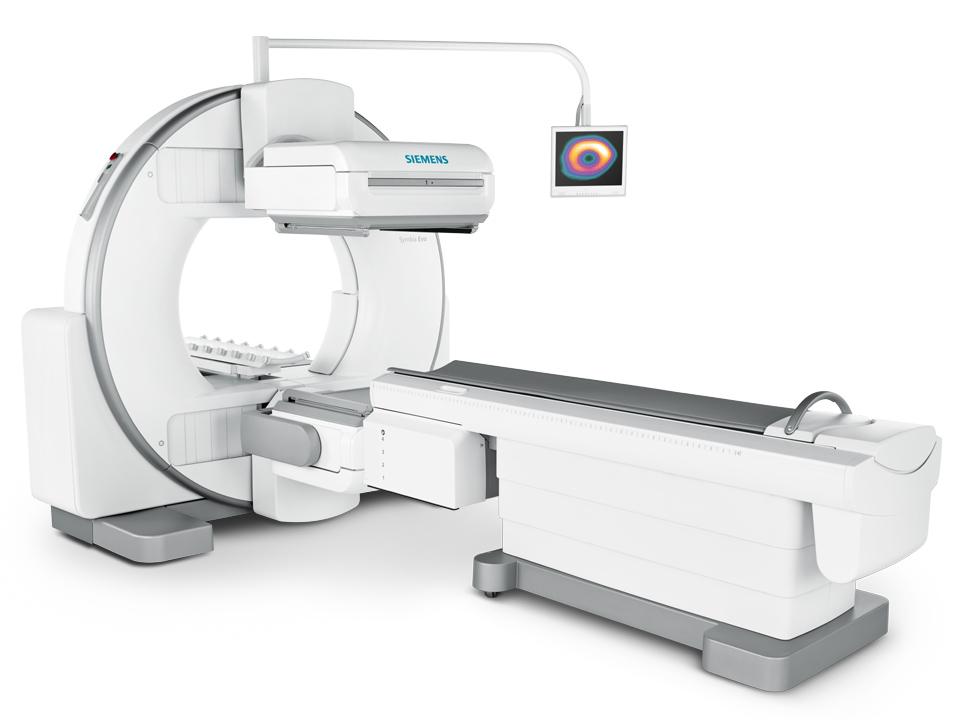
How does a SPECT scan work?
A SPECT scan involves two main steps. Your provider will:
- Give you a radiotracer. A radiotracer is a radioactive substance that helps highlight certain areas in your body on an imaging test. Providers usually inject radiotracers into a vein. But in some cases, you may need to swallow or inhale the substance. As the radiotracer moves through your body, it accumulates in certain areas. This gives your provider information about how your organs and tissues function. Radiotracers are safe. They contain very minimal amounts of radiation — about the same amount you get from a regular X-ray. They don’t contain dyes or cause side effects and they leave your body within 24 hours.
- Take pictures with a gamma camera. A gamma camera (nuclear camera) uses specialized imaging techniques to look for the radiotracers in your body. You can think of a gamma camera as a radiation detector. It doesn’t emit radiation, but it finds radioactive energy (in this case, the radiotracer) inside your body. The gamma camera takes pictures of your organs, bones and tissues, and tells your provider how well they’re working.
How do I prepare for a SPECT scan?
Your provider will give you specific instructions for your situation. In general, you should wear comfortable clothing and leave jewelry, watches and other metal items at home.
You'll receive a radioactive substance through an intravenous (IV) infusion into a vein in your arm. The tracer dose is very small, and you may feel a cold sensation as it enters your body. You may be asked to lie quietly in a room for 20 minutes or more before your scan while your body absorbs the radioactive tracer. In some cases, you may need to wait several hours or, rarely, several days between the injection and your SPECT scan.
Your body's more active tissues will absorb more of the radioactive substance. For instance, during a seizure, the area of your brain causing the seizure may hold on to more of the radioactive tracer. This can pinpoint the area of the brain causing your seizures.
What to expect during a SPECT scan
Once your body absorbs the radiotracer, your provider will walk you to a room with a SPECT machine. You’ll lie down on a table (usually on your back) while the scanner rotates around you. The SPECT machine will take pictures of the structures inside your body. Then, it’ll send the information to a computer, which will create detailed 3D images.
A SPECT scan usually takes about 30 minutes to complete. It may take longer if your provider needs to take pictures of other areas, can take up to 3 hours.
What to expect after the test
Most of the radioactive tracer leaves your body through your urine within a few hours after your SPECT scan. You may be told to drink more fluids, such as juice or water, after your SPECT scan. This helps flush the tracer from your body. Your body breaks down the remaining tracer over the next few days. Once the scan is completed, you can usually leave and resume your daily activities right away.
What are the advantages of a SPECT scan?
SPECT scans:
- Are safe for people with pacemakers and
other cardiovascular implantable electronic devices
(CIEDs).
- Can find issues that other imaging methods can’t detect.
- Can tell your provider how well your organs function.
Are there any risks or possible complications?
SPECT scans:
- Are safe for people with pacemakers and other cardiovascular implantable electronic devices (CIEDs).
- Can find issues that other imaging methods can’t detect.
- Can tell your provider how well your organs function.
Your healthcare provider will use a small amount of radiation during a SPECT scan. The exact dosage depends on factors like your size and what type of radiotracer your provider uses.
Risks from radiation during a SPECT scan are minimal. Talk to your healthcare provider if you have questions or concerns about your radiation exposure.
Risks
For most people, SPECT scans are safe. If you have an injection or infusion of radioactive tracer, you may experience:
- You’re pregnant or nursing: The tests use a low dose of radiation, which is not recommended for pregnant women.6 If you’re breastfeeding, you may be required to wait a certain amount of time before nursing to allow your body time to excrete the radioactive tracer.
- Discomfort During Injection: Some individuals may feel mild discomfort or notice slight bruising at the injection site.
- You're allergic to the tracer: Though unusual, this kind of allergy is possible, and you shouldn't have the scan if you have a known allergy to the tracer. If you have an allergic reaction while undergoing the scan, know that the healthcare professionals around you are equipped to handle the situation.
Be sure to tell your healthcare team or radiation technologist if there's a possibility you're pregnant or if you're breastfeeding.
Risks of radiation
Your healthcare team uses a small amount of radiation to perform a SPECT scan, and the test is not associated with any long-term health risks. Talk to someone on your team if you're concerned about your exposure to radiation during a SPECT scan.
When should I know the results of my SPECT scan?
A radiologist or healthcare specialist with advanced training in nuclear medicine will study the results of your SPECT scan and send them to your healthcare team. Pictures from your scan may show colors that tell your team what areas of your body absorbed more of the radioactive tracer and which areas absorbed less. For instance, a brain SPECT image might show a lighter color where brain cells are less active and darker colors where brains cells are more active. Some SPECT images show shades of gray, rather than colors. Ask your healthcare team how long to expect to wait for your results.
You should get the results of your SPECT scan back in about one week. After your appointment, a radiologist will interpret the images captured during your scan. Then they’ll create a report of their findings to share with the provider who ordered the test. Your provider will talk with you about your results and determine any appropriate next steps.
Additional Details
What is the difference between a SPECT and CT scan?
A CT (computed tomography) scan uses radiation to take detailed pictures of the structures inside your body. The main goal of a CT scan is to look at your anatomy. It shows the size and location of organs, bones and tissues.
A SPECT scan involves injecting, ingesting or inhaling a radiotracer before taking images. The main goal of a SPECT scan is to look at your physiology — how the radiotracer behaves once it’s inside your body. This is helpful for determining how your organs and tissues function.
In some cases, a healthcare provider may combine CT and SPECT technology to get even more detailed information. Some scanners can take both types of images at the same time.
What is the difference between a SPECT scan and an MRI?
Magnetic resonance imaging (MRI) uses a large magnet and radio waves to take pictures of the structures inside your body. Like a CT scan, MRI can tell you a lot about your physical anatomy — but it can’t tell you how your anatomy functions.
A SPECT scan, on the other hand, shows how your organs and tissues work. After your provider injects the radiotracer, the substance moves through your body and accumulates in certain areas. How the radiotracer behaves can tell your provider whether your organs, bones and tissues function as they should.
Food and Drink
Your healthcare team will let you know if the scan requires you to avoid certain foods or drinks.
For example, if you have a SPECT scan for cardiac reasons, you may need to avoid caffeine for several hours before the test.
Cost and Health Insurance
Your insurance may require prior authorization in order to cover your SPECT scan. Be sure to check with the company on whether and to what extent the scan is covered so you'll know what, if any, costs you'll need to cover.
SPECT scans, without insurance coverage, can cost over $1,300 to $4,000 or more.
One Final Note..
Providers can use SPECT scans to evaluate any area of your body. But this type of imaging is most common for detecting heart, brain and bone conditions. Talk to your healthcare provider to learn more about SPECT scans and whether you need one.
Find me on Social Media


 |
Don't forget to bookmark me to see updates.. Copyright © 2000 - 2025 K.
Kerr |