- Main Page
- A1C Test
- Advance Directives
- Age on other Planets
- Aliens
- American Flag
- Annuals
- Anxiety
- Aortic Aneurysm
- Apple Cider Vinegar
- Arrhythmia
- Atrial Fibrillation
- Avoiding Scams
- Awareness Ribbons
- Bamboo
- Banana Tree, Grand Nain
- Banana Tree, Ice Cream
- Banana Tree, Zebrina Rojo
- Beekeeping
- Benign P P Vertigo
- Birth Month
- Blood Tests
- Blood Types
- Body Mass Index - BMI
- BMI Calculator
- Boogaloo
- Bookmarks
- Boot Anatomy
- Boot Fit Guide
- Boot Glossary
- Boot Leathers
- Boot Makers
- Boot Retailers
- Boot Styles - Western
- Boot Toes & Heels - Western
- Boot Toes & Heels - Work
- Bronchitis
- Candle Colors
- Carbohydrates
- Cardiac Catheterization
- Cardiovascular Disease
- CGM's
- Chakras
- Chinese Zodiac
- Cholesterol
- Christmas Tree
- Color Codes Chart
- C.O.P.D.
- Coronary Artery Disease
- Country Stars
- Cowboy Hat Etiquette
- Cowboy Hat Sizing
- C.P.A.P.
- Credit Score Checkers
- Crystals & Gems
- CT scan
- Degenerative Disk Disease
- Depression
- Diabetes Info.
- Diabetes Facts
- Diabetes - Pre
- Diabetes - Type 1
- Diabetes - Type 2
- Diabetes - Type 3c
- Diabetes - Gestational
- Diabetes Care
- Diabetes Care Team
- Diabetes Terms
- Diabetes Treatment
- Diabetes & Fruits
- Diabetes & Veg's
- Diet - Boiled Egg
- Diet - DASH
- Diet - Fat Burning
- Diet - Mediterranean
- Diet - Military
- Disability
- Do Not Resuscitate
- Dream Catchers
- Dupixent®
- Echocardiogram
- Electrocardiogram
- Emphysema
- Epsom Salt
- Eye Teasers
- Fairies
- Farxiga®
- Flower Astrology
- Fonts
- Foods To Regrow
- Friend
- Funny Things
- Fun Stuff
- Glycemic Index
- Gout
- Growing Blueberries
- Halloween
- Halloween Treats
- Headaches
- Health Info. Lines
- Heart Attack
- Heart Disease - Other
- Heart Failure
- Heart Tests
- Hello!!
- Herbal Codes
- Herbal Medicine
- Herb & Oils Uses
- Herniated disk
- Home Remedies
- House Plants
- Humalog®
- Hydrogen Peroxide
- Hyperglycemia
- Hypoglycemia
- Hyperkalemia
- Hypokalemia
- Hypertension
- Hypotension
- Important Numbers
- Insomnia
- Insulin
- Juice Recipes
- Karma
- Kidney Cysts
- Kidney Disease
- Kinds of Tea
- Lantus®
- Lemon Cleanse
- Logger vs Lineman
- Macaroni!!
- Medicare
- Mental Health
- MO HealthNet
- Moon Phases
- Mounjaro®
- MRI Scan
- My Athletic Shoes
- My Boots & Spurs
- My Cowboy Hats
- Myelography
- Mystical Unicorn
- Nasal Polyps
- Natal Astrology Chart
- Never Forget
- Nuclear Medicine
- Nutrition - Adults
- Nutrition - Adults, Older
- Nutrition - Kids
- Obesity
- One Little Rose
- Orchid Growing
- Orchid Sources
- Pagan Humor
- Pagans vs.Wiccans
- Parking Spaces
- PayPal.Me
- Pentagram vs. Pentacle
- Perennials
- Peripheral Artery Disease
- PET/CT Scan
- PET Scan
- Phobias A-Z
- Plant Care
- Plant Zone Map
- Potassium
- Propagating Plants
- Prurigo Nodularis
- Psychic Gifts
- PVC's
- Quit Smoking
- Recipes I like
- Red Yeast Rice
- Roses
- Runes
- Sadie & Beethoven
- Salt & Sodium
- Salt Water Flush
- Sciatica
- Service Animals
- Shape Shifters
- Sleep Apnea
- Sleep Disorders
- Sleep Studies
- Smile
- SPECT Scan
- Speed Test
- Spices You Need
- Spices I Have
- Spinal Stenosis
- Stents
- Steel Toe vs. Comp. Toe
- Stress Test - Exercise
- Stress Test - Nuclear
- Sugars - Sweeteners
- Superstitions
- Symbols
- Tarot
- The Ten Commandments
- Tools of the Craft
- Top Expensive Movies
- Top Modern Westerns
- Top 100 Westerns
- Toyota Yaris 2008
- Toyota Yaris 2012
- Trazodone
- Tree, Calamondin Orange
- Tree, Lemon (Meyer)
- Tree, Lime
- Tree Signs
- Ultrasound
- US Bill of Rights
- US Constitution
- US Declaration of Independence
- Vaccines by Age
- Vaccines 0-6 yrs
- Vaccines 7-18 yrs
- Vaccines 19 and up
- Ventricular Fibrillation
- Vertigo
- Vital Records
- Vital Signs
- Vitamin B12
- Vitamin C
- Vitamin D
- Vitamin E
- Vitamin K
- Vitamins & Minerals
- Water Therapy
- Weight on other Planets
- Wiccan Rede
- X-Rays
- Yin / Yang
- Zodiac Signs
Needed to read PDF's

Magnetic Resonance Imaging
(MRI)
Overview
Magnetic resonance imaging (MRI) is a medical imaging technique that uses a magnetic field and computer-generated radio waves to create detailed images of the organs and tissues in your body.
Most MRI machines are large, tube-shaped magnets. When you lie inside an MRI machine, the magnetic field inside works with radio waves and hydrogen atoms in your body to create cross-sectional images — like slices in a loaf of bread.
The MRI machine also can produce 3D images that can be viewed from different angles.
What is an MRI?
An MRI (magnetic resonance imaging) scan is a painless test that produces very clear images of the organs and structures inside your body. MRI uses a large magnet, radio waves and a computer to produce these detailed images. It doesn’t use X-rays (radiation).
Because MRI doesn’t use X-rays or other radiation, it’s the imaging test of choice when people will need frequent imaging for diagnosis or treatment monitoring, especially of their brain.
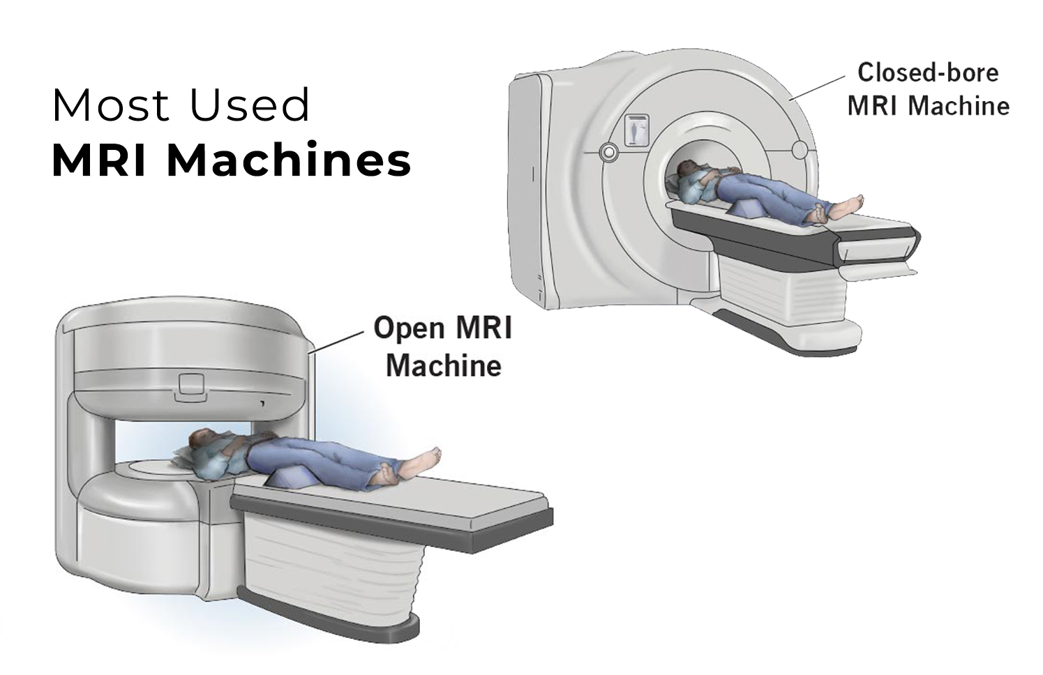
What is an open MRI?
An open (or “open bore”) MRI refers to the type of machine that takes the images. Typically, an open MRI machine has two flat magnets positioned over and under you with a large space between them for you to lie. This allows for open space on two sides and alleviates much of the claustrophobia many people experience with closed-bore MRI machines.
However, open MRIs don’t take as clear images as closed-bore MRI machines. Closed-bore MRI machines have a ring of magnets that forms an open hole or tube in the middle where you’d lie to get the images. Closed-bore MRIs are narrow with tight head-to-ceiling space. This can cause anxiety and discomfort for some people, but these MRI machines take the best quality images.
If you’re nervous about your MRI scan or have a fear of closed spaces, talk to your healthcare provider. If needed, your provider will discuss options for sedatives (medicines to make you feel relaxed) or even anesthesia if necessary.
What is an MRI with contrast?
Some MRI exams use an injection of contrast material. The contrast agent contains gadolinium, which is a rare earth metal. When this substance is present in your body, it alters the magnetic properties of nearby water molecules, which enhances the quality of the images. This improves the sensitivity and specificity of the diagnostic images.
Contrast material enhances the visibility of the following:
- Tumors.
- Inflammation.
- Infection.
- Blood supply to certain organs.
- Blood vessels.
If your MRI requires a contrast material, a healthcare provider will insert an intravenous catheter (IV line) into a vein in your hand or arm. They’ll use this IV to inject the contrast material.
Contrast materials are safe drugs. Side effects ranging from mild to severe do occur, but severe reactions are very rare.
What’s the difference between an MRI scan and a CT scan?
Magnetic resonance imaging (MRI) uses magnets, radio waves and a computer to create images of the inside of your body, whereas computed tomography (CT) uses X-rays and computers.
Healthcare providers often prefer to use MRI scans instead of CT scans to look at the non-bony parts or soft tissues inside your body. MRI scans are also safer since they don’t use the damaging ionizing radiation of X-rays.
MRI scans also take much clearer pictures of your brain, spinal cord, nerves, muscles, ligaments and tendons than regular X-rays and CT scans.
However, not everyone can undergo an MRI. The magnetic field of MRI can displace metal implants or affect the function of devices such as pacemakers and insulin pumps. If this is the case, a CT scan is the next best option.
MRI scanning is usually more expensive than X-ray imaging or CT scanning.
What does an MRI show?
Magnetic resonance imaging (MRI) produces detailed images of the inside of your body. Healthcare providers can “look at” and evaluate several different structures inside your body using MRI, including:
- Your brain and surrounding nerve tissue.
- Organs in your chest and abdomen, including your heart, liver, biliary tract, kidneys, spleen, bowel, pancreas and adrenal glands.
- Breast tissue.
- Your spine and spinal cord.
- Pelvic organs, including your bladder and reproductive organs (uterus and ovaries in females and the prostate gland in males).
- Blood vessels.
- Lymph nodes.
Why it's done
MRI is a noninvasive way for a medical professional to examine your organs, tissues and skeletal system. It produces high-resolution images of the inside of the body that help diagnose a variety of conditions.
MRI of the brain and spinal cord
MRI is the most frequently used imaging test of the brain and spinal cord. It's often performed to help diagnose:
- Aneurysms of cerebral vessels.
- Conditions of the eye and inner ear.
- Multiple sclerosis.
- Spinal cord conditions.
- Stroke.
- Tumors.
- Brain injury from trauma.
A special type of MRI is the functional MRI of the brain, also known as fMRI. It produces images of blood flow to certain areas of the brain. Functional MRI can be used to examine the brain's anatomy and show which parts of the brain are handling critical functions, language and movements. This information can help guide decisions when considering someone for brain surgery.
Functional MRI also can check for damage from a head injury or from conditions such as Alzheimer's disease.
MRI of the heart and blood vessels
MRI that focuses on the heart or blood vessels can check:
- Size and function of the heart's chambers.
- Thickness and movement of the walls of the heart.
- Extent of damage caused by heart attacks or heart disease.
- Structural problems in the aorta, such as aneurysms or dissections.
- Inflammation or blockages in the blood vessels.
MRI of other internal organs
MRI can check for tumors or other irregularities in many organs in the body, including the following:
- Liver and bile ducts.
- Kidneys.
- Spleen.
- Pancreas.
- Uterus.
- Ovaries.
- Prostate.
MRI of bones and joints
MRI can help look for:
- Joint issues caused by traumatic or repetitive injuries, such as torn cartilage or ligaments.
- Disk problems in the spine.
- Bone infections.
- Tumors of the bones and soft tissues.
MRI of the breasts
MRI can be used with mammography to detect breast cancer, particularly in people who have dense breast tissue or who might be at high risk of the disease.
Risks
Because MRI uses powerful magnets, the presence of metal in your body can be a safety hazard if attracted to the magnet. Even if not attracted to the magnet, metal objects can distort the MRI images. Before having an MRI exam, you'll likely complete a questionnaire that includes whether you have metal or electronic devices in your body.
Unless the device you have is certified as MRI safe, you might not be able to have an MRI.
Devices include:
- Metallic joint prostheses.
- Artificial heart valves.
- An implantable heart defibrillator.
- Implanted drug infusion pumps.
- Implanted nerve stimulators.
- A pacemaker.
- Metal clips.
- Metal pins, screws, plates, stents or surgical staples.
- Cochlear implants.
- A bullet, shrapnel or any other type of metal fragment.
- Intrauterine device.
If you have tattoos or permanent makeup, ask whether it might affect your MRI. Some of the darker inks contain metal.
Before you schedule an MRI, tell your doctor if you think you're pregnant. The effects of magnetic fields on an unborn baby aren't well understood. An alternative exam may be recommended, or the MRI may be postponed. Also tell your doctor if you're breastfeeding, especially if you're to receive contrast material during the procedure.
It's also important to discuss kidney or liver problems with your doctor and the technologist, because problems with these organs might limit the use of injected contrast agents during your MRI scan.
What do I need to do to prepare for an MRI?
The magnetic resonance imaging (MRI) scanner uses strong magnets and radio wave signals that can cause heating or possible movement of some metal objects in your body. This could result in health and safety issues. It could also cause some implanted electronic medical devices to malfunction.
If you have metal-containing objects or implanted medical devices in your body, your healthcare provider needs to know about them before your MRI scan. Certain implanted objects may require additional scheduling arrangements and special instructions. Other items don’t require special instructions but may require an X-ray to check on the exact location of the object before your exam.
Please tell your provider and MRI technologist if you have any of the following:
- Heart pacemaker or defibrillator.
- Electronic or implanted stimulators or devices, including deep brain stimulators, vagus nerve stimulators, bladder stimulators, spine stimulators, neurostimulators and implanted electrodes or wires.
- Metallic joint prostheses.
- Cochlear implant or other ear implants.
- Implanted drug pumps, such as those that pump narcotic/pain medications or drugs to treat spasticity.
- Programmable shunt.
- Aneurysm clips and coils.
- Stents not located in your heart.
- Filters, such as blood clot filters.
- Metal fragments in your body or eye, such as bullets, shrapnel, metal pieces or shavings.
You won’t be able to wear the following devices during your MRI. Please coordinate your MRI appointment with the day you need to change your patch or device.
- Continuous glucose monitor (CGM).
- Insulin pump.
- Medication patches.
In addition, tell your provider if you:
- Are pregnant.
- Are not able to lie on your back for 30 to 60 minutes.
- Have claustrophobia (fear of enclosed or narrow spaces).
Leave all jewelry and other accessories at home or remove them before your MRI scan. Metal and electronic items aren’t allowed in the exam room because they can interfere with the magnetic field of the MRI unit, cause burns or become harmful projectiles. These items include:
- Jewelry, watches, credit cards and hearing aids — all of which can be damaged.
- Pins, metal hair accessories, under wire bras and metal zippers, which can distort MRI images.
- Removable dental work, such as dentures.
- Pens, pocketknives and eyeglasses.
- Body piercings.
- Cell phones, electronic watches and tracking devices.
What you can expect
During the test
The MRI machine looks like a long narrow tube that is open on both ends. You lie down on a movable table that slides into the opening of the tube. A technologist monitors you from another room. You can talk with the technologist by microphone.
If you have a fear of enclosed spaces, called claustrophobia, you might receive a drug to help you feel sleepy and less anxious. Most people get through the exam without difficulty.
The MRI machine creates a strong magnetic field around you, and radio waves are directed at your body. The procedure is painless. You don't feel the magnetic field or radio waves, and there are no moving parts around you.
During the MRI scan, the internal part of the magnet produces repetitive tapping, thumping and other noises. Wearing earplugs or having music playing can help block the noise.
In some cases, a contrast material, typically gadolinium, will be injected through an intravenous (IV) line into a vein in a hand or arm. The contrast material helps make certain details clearer. Gadolinium rarely causes allergic reactions.
An MRI exam can last anywhere from 15 minutes to more than an hour. You must hold still because movement can blur the images.
During a functional MRI exam, you might be asked to perform a few small tasks — such as tapping your thumb against your fingers, rubbing a block of sandpaper or answering simple questions. This helps pinpoint the portions of your brain that control these actions.
What are the side effects of MRI contrast?
On very rare occasions, some people who have contrast material for their MRI experience side effects, including:
- Nausea.
- Headache.
- Pain at the site of the injection.
It’s very rare to experience hives, itchy eyes or other signs of an allergic reaction to the contrast material. If you have allergic symptoms, tell the technologist. A healthcare provider will be available to provide immediate medical care.
Nephrogenic systemic fibrosis (NSF), which causes thickening of your skin, organs and other tissues, is a rare complication in people with kidney disease that undergo an MRI with contrast material. Because of this, people with severe kidney disease may not be able to have gadolinium-based contrast material for their MRI.
There’s evidence that tiny traces of gadolinium may stay in different organs of your body after contrast-enhanced MRI. While there are no known negative effects from this, your provider may take gadolinium retention into account when selecting a contrast agent.
After the test
If you didn’t have a sedative drug for the MRI scan, no recovery period is necessary. You can go home and resume your normal activities. If you had sedative drugs for the exam, you’ll need to recover from the effects of them before you can go home. You may need to have someone else drive you home.
Results
A doctor specially trained to interpret MRI scans, called a radiologist, will look over the images from your scan and report the findings to your doctor. Your doctor will discuss important findings and next steps with you.
An MRI is a very useful tool for helping your doctors see images of the inside of your body, including tissue that can't be seen on a conventional x-ray.
Before your exam, it's very important to fill out the safety screening form carefully. MRI is safe and painless. But metal in the scanner can cause serious safety problems or reduce the quality of the images.Your health care team needs to know about any metal in your body, even a small shard of metal from an accident. Fillings, bridges, and other dental work typically do not pose a problem. But other metal that has been put into your body might prevent you from having an MRI. That includes some pacemakers, clips for treating aneurysms, and other devices with metal in them.
A nurse may review your health history before your exam. You may be given medications or contrast dye or have blood drawn. Be sure to tell the nurse if you're pregnant, have an allergy to contrast dye, or have kidney or liver problems. You may not wear clothing with snaps or zippers in the scanner. You will be asked to wear a gown. Do not wear any jewelry or bring anything metal into the scanner, including a hearing aid.
An MRI machine uses a powerful magnet to make images of your body. Unlike a CT scan, it does not use x-rays or other radiation. You will be given earplugs. The scanner makes a loud noise when it's operating.
A device called a coil may be put on or around the area to be scanned to help capture the images. You will also be given a squeeze ball to hold. You can use this to signal the technologist any time you need something. The MRI is controlled from a nearby room. You will be closely observed throughout the procedure.
A series of scans are taken with a brief pause between each. You may hear different noises as different scans are taken. It's normal for the noise to be very loud. You need to remain still when the scan is being taken.
People are typically in the scanner from 30 to 50 minutes, depending on the images to be taken. A complex examination can take longer. If you are concerned about being in the scanner for this length of time, talk to your physician and the technologist. They can help you with some tips for staying comfortable.
If you need to be removed from the scanner, this can be done very quickly. The ends of the scanner are always open.
After your exam, the images will be reviewed by your radiologist. He or she will send a report to the health care provider who ordered the test. Ask your health care provider any questions you have about your MRI.
Find me on Social Media


 |
Don't forget to bookmark me to see updates.. Copyright © 2000 - 2025 K.
Kerr |