- Main Page
- A1C Test
- Advance Directives
- Age on other Planets
- Aliens
- American Flag
- Annuals
- Anxiety
- Aortic Aneurysm
- Apple Cider Vinegar
- Arrhythmia
- Atrial Fibrillation
- Avoiding Scams
- Awareness Ribbons
- Bamboo
- Banana Tree, Grand Nain
- Banana Tree, Ice Cream
- Banana Tree, Zebrina Rojo
- Beekeeping
- Benign P P Vertigo
- Birth Month
- Blood Tests
- Blood Types
- Body Mass Index - BMI
- BMI Calculator
- Boogaloo
- Bookmarks
- Boot Anatomy
- Boot Fit Guide
- Boot Glossary
- Boot Leathers
- Boot Makers
- Boot Retailers
- Boot Styles - Western
- Boot Toes & Heels - Western
- Boot Toes & Heels - Work
- Bronchitis
- Candle Colors
- Carbohydrates
- Cardiac Catheterization
- Cardiovascular Disease
- CGM's
- Chakras
- Chinese Zodiac
- Cholesterol
- Christmas Tree
- Color Codes Chart
- C.O.P.D.
- Coronary Artery Disease
- Country Stars
- Cowboy Hat Etiquette
- Cowboy Hat Sizing
- C.P.A.P.
- Credit Score Checkers
- Crystals & Gems
- CT scan
- Degenerative Disk Disease
- Depression
- Diabetes Info.
- Diabetes Facts
- Diabetes - Pre
- Diabetes - Type 1
- Diabetes - Type 2
- Diabetes - Type 3c
- Diabetes - Gestational
- Diabetes Care
- Diabetes Care Team
- Diabetes Terms
- Diabetes Treatment
- Diabetes & Fruits
- Diabetes & Veg's
- Diet - Boiled Egg
- Diet - DASH
- Diet - Fat Burning
- Diet - Mediterranean
- Diet - Military
- Disability
- Do Not Resuscitate
- Dream Catchers
- Dupixent®
- Echocardiogram
- Electrocardiogram
- Emphysema
- Epsom Salt
- Eye Teasers
- Fairies
- Farxiga®
- Flower Astrology
- Fonts
- Foods To Regrow
- Friend
- Funny Things
- Fun Stuff
- Glycemic Index
- Gout
- Growing Blueberries
- Halloween
- Halloween Treats
- Headaches
- Health Info. Lines
- Heart Attack
- Heart Disease - Other
- Heart Failure
- Heart Tests
- Hello!!
- Herbal Codes
- Herbal Medicine
- Herb & Oils Uses
- Herniated disk
- Home Remedies
- House Plants
- Humalog®
- Hydrogen Peroxide
- Hyperglycemia
- Hypoglycemia
- Hyperkalemia
- Hypokalemia
- Hypertension
- Hypotension
- Important Numbers
- Insomnia
- Insulin
- Juice Recipes
- Karma
- Kidney Cysts
- Kidney Disease
- Kinds of Tea
- Lantus®
- Lemon Cleanse
- Logger vs Lineman
- Macaroni!!
- Medicare
- Mental Health
- MO HealthNet
- Moon Phases
- Mounjaro®
- MRI Scan
- My Athletic Shoes
- My Boots & Spurs
- My Cowboy Hats
- Myelography
- Mystical Unicorn
- Nasal Polyps
- Natal Astrology Chart
- Never Forget
- Nuclear Medicine
- Nutrition - Adults
- Nutrition - Adults, Older
- Nutrition - Kids
- Obesity
- One Little Rose
- Orchid Growing
- Orchid Sources
- Pagan Humor
- Pagans vs.Wiccans
- Parking Spaces
- PayPal.Me
- Pentagram vs. Pentacle
- Perennials
- Peripheral Artery Disease
- PET/CT Scan
- PET Scan
- Phobias A-Z
- Plant Care
- Plant Zone Map
- Potassium
- Propagating Plants
- Prurigo Nodularis
- Psychic Gifts
- PVC's
- Quit Smoking
- Recipes I like
- Red Yeast Rice
- Roses
- Runes
- Sadie & Beethoven
- Salt & Sodium
- Salt Water Flush
- Sciatica
- Service Animals
- Shape Shifters
- Sleep Apnea
- Sleep Disorders
- Sleep Studies
- Smile
- SPECT Scan
- Speed Test
- Spices You Need
- Spices I Have
- Spinal Stenosis
- Stents
- Steel Toe vs. Comp. Toe
- Stress Test - Exercise
- Stress Test - Nuclear
- Sugars - Sweeteners
- Superstitions
- Symbols
- Tarot
- The Ten Commandments
- Tools of the Craft
- Top Expensive Movies
- Top Modern Westerns
- Top 100 Westerns
- Toyota Yaris 2008
- Toyota Yaris 2012
- Trazodone
- Tree, Calamondin Orange
- Tree, Lemon (Meyer)
- Tree, Lime
- Tree Signs
- Ultrasound
- US Bill of Rights
- US Constitution
- US Declaration of Independence
- Vaccines by Age
- Vaccines 0-6 yrs
- Vaccines 7-18 yrs
- Vaccines 19 and up
- Ventricular Fibrillation
- Vertigo
- Vital Records
- Vital Signs
- Vitamin B12
- Vitamin C
- Vitamin D
- Vitamin E
- Vitamin K
- Vitamins & Minerals
- Water Therapy
- Weight on other Planets
- Wiccan Rede
- X-Rays
- Yin / Yang
- Zodiac Signs
Needed to read PDF's

Positron Emission Tomography
(PET)
Overview
A positron emission tomography (PET) scan is an imaging test that can help reveal the metabolic or biochemical function of your tissues and organs. The PET scan uses a radioactive drug called a tracer to show both typical and atypical metabolic activity. A PET scan can often detect the atypical metabolism of the tracer in diseases before the disease shows up on other imaging tests, such as computerized tomography (CT) and magnetic resonance imaging (MRI).
The tracer is most often injected into a vein within your hand or arm. The tracer will then collect into areas of your body that have higher levels of metabolic or biochemical activity. This often pinpoints the location of the disease. The PET images are typically combined with CT or MRI and are called PET-CT or PET-MRI scans.
What is a PET scan?
A positron emission tomography (PET) scan is an imaging test that produces images of your organs and tissues at work. The test uses a safe, injectable radioactive chemical called a radiotracer and a device called a PET scanner.
The scanner detects diseased cells that absorb large amounts of the radiotracer, which indicates a potential health problem.
Healthcare providers frequently use PET scans to help diagnose cancer and assess cancer treatment. They can also assess certain heart and brain issues with the scan.
What’s the difference between a PET scan, CT scan and MRI?
Computed tomography (CT) scans use X-rays. Magnetic resonance imaging (MRI) scans use magnets and radio waves. Both produce still images of organs and body structures.
PET scans use a radioactive tracer to show how an organ is functioning in real time. PET scan images can detect cellular changes in organs and tissues earlier than CT and MRI scans. Your healthcare provider may perform a PET scan and CT scan at the same time (PET-CT). This combination test produces 3D images that allow for a more accurate diagnosis.
Some hospitals now use a hybrid PET/MRI scan. This new technology creates extremely high-contrast images. Providers mainly use this type of scan for diagnosing and monitoring cancers of the soft tissues (brain, head and neck, liver and pelvis).
Why it's done
A PET scan is an effective way to help discover a variety of conditions, including cancer, heart disease and brain disorders. Your health care provider can use this information to help diagnose, monitor or treat your condition.
Cancer
Cancer cells show up as bright spots on PET scans because they have a higher metabolic rate than do typical cells. PET scans may be useful in:
- Detecting cancer.
- Revealing whether your cancer has spread.
- Checking whether a cancer treatment is working.
- Finding a cancer recurrence.
scans must be interpreted carefully because noncancerous conditions can look like cancer. Also, some cancers do not appear on PET scans. Many types of solid tumors can be detected by PET-CT and PET-MRI scans, including:
- Brain.
- Breast.
- Cervical.
- Colorectal.
- Esophageal.
- Head and neck.
- Lung.
- Lymphatic system.
- Pancreatic.
- Prostate.
- Skin.
- Thyroid.
Heart disease
A positron emission tomography (PET) scan of the heart is an imaging test that uses specialized dye to allow your doctor to view problems with your heart.
The dye contains radioactive tracers, which concentrate on areas of the heart that may be injured or diseased. Using a PET scanner, your doctor can spot these areas of concern.
A heart PET scan is typically an outpatient procedure, meaning you will not have to stay at the hospital overnight. This is typically a same-day procedure.
Your doctor may order a heart PET scan if you’re experiencing symptoms of heart trouble.
Symptoms of heart trouble include:
- irregular heartbeat (arrhythmia)
- pain in your chest
- tightness in your chest
- trouble breathing
- weakness
- profuse sweating
Your doctor may also order a heart PET scan if other heart tests, such as an echocardiogram (ECG) or cardiac stress test, don’t provide your doctor with enough information. A heart PET scan can also be used to track the effectiveness of heart disease treatments.
Brain disorders
Glucose is the main fuel of the brain. During PET scans, tracers are “attached” to compounds such as glucose. By detecting radioactive glucose, the PET scan can show which areas of the brain are using glucose at the highest rates.
When a specialist interprets the scan, they can see how the brain is working and check for any irregularities.
PET scans are used to help diagnose and manage many CNS disorders, including:
- Alzheimer’s disease
- depression
- epilepsy
- head trauma
- Parkinson’s disease
When would I need a PET scan?
In general, a PET scan can measure vital functions, such as blood flow, oxygen use and blood sugar (glucose) metabolism. It can also identify organs and tissues that aren’t working as they should.
If your healthcare provider suspects you may have cancer, they’ll likely recommend a PET scan, which can detect cancer and/or make a diagnosis.
If you’ve already been diagnosed with cancer, your provider may recommend more than one PET scan throughout your treatment to:
- Determine whether the cancer has spread in your body (metastasized).
- Assess the effectiveness of treatment.
- Determine if the cancer has returned after treatment (recurred).
- Evaluate the prognosis (outlook) of the cancer.
If you’re having heart issues, your provider may recommend a PET scan to:
- Determine the effects of a heart attack on areas of your heart.
- Identify areas of the heart muscle that would benefit from angioplasty or coronary artery bypass surgery.
If you’re experiencing neurological symptoms, your provider may recommend a PET scan to evaluate possible brain abnormalities, such as tumors, seizures and other central nervous system conditions.
Test Details
How does a PET scan work?
A PET scan is a type of nuclear medicine imaging. Nuclear medicine uses small and safe amounts of radioactive material, called radiotracers, given through an IV.
Unlike other imaging techniques, PET scans focus on processes and molecular activity within your body. This gives them the potential to find disease in its earliest stages.
Diseased cells in your body absorb more of the radiotracer than healthy ones do. These are called “hot spots.” The PET scanner detects this radiation and produces images of the affected tissue. A PET/CT scan combines X-ray images from a CT scan with PET scan images.
How do I prepare for a PET scan?
PET scans are an outpatient procedure, which means you go home the same day. Your healthcare provider will give you detailed instructions on how to prepare for the scan. In general, you should:
- Make sure your provider has a current list of all medications, vitamins and supplements you take, as well as any allergies you have.
- Alert your provider if you think you could be pregnant or if you’re breastfeeding.
- Not eat anything for six hours before the test. Your healthcare provider may change this direction if you have diabetes.
- Drink only water.
- Avoid caffeine for 24 hours before the test if you’re being tested for a heart problem.
- Wear comfortable clothes and leave metal accessories, such as jewelry, eyeglasses, dentures and hairpins at home.
- Tell your healthcare provider if being in an enclosed space makes you anxious. You may be able to take a mild sedative to help you relax during the procedure.
What should I expect during a PET scan?
You can expect the following during a PET scan:
- You’ll receive an IV injection of a radiotracer that contains a safe amount of a radioactive drug. The most commonly used radiotracer is fluorodeoxyglucose (FDG).
- You’ll sit in a chair for about an hour while the radiotracer moves through your bloodstream and gets absorbed by your organs and tissues. Too much activity can send the radiotracer to areas of your body that your healthcare provider isn’t testing. You won’t be able to feel the radiotracer.
- If you’re getting a PET/CT scan, you may also get an IV injection of a contrast dye. This dye helps produce sharper CT images.
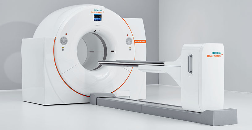
- You’ll lie on an exam table that slides in and out of the PET/CT scanner. This scanner is shaped like a doughnut. The doughnut or tunnel opening is about 30 inches in diameter.
- During the scan, which usually takes about 30 minutes, you must remain still. Movement can blur the images.
- You’ll hear buzzing and clicking sounds as the scanner takes images.
- A technologist will review the scans before you leave to ensure the images are in focus.
How long does a PET scan take?
The entire PET scan process takes about two hours.
It can take up to 60 minutes for your body to absorb the injected radiotracer. During this time, you’ll need to sit quietly and limit your movements. The actual PET scan takes about 30 minutes. After the test, you’ll need to wait while the technologist reviews the scans to ensure the images are clear.
What will I experience during and after the procedure?
Except for intravenous injections, most nuclear medicine procedures are painless. Reports of significant discomfort or side effects are rare.
You will feel a slight pin prick when the technologist inserts the needle into your vein for the intravenous line. You may feel a cold sensation moving up your arm during the radiotracer injection. Generally, there are no other side effects.
PET scans only use radiotracer injections.
With some procedures, the technologist may place a catheter into your bladder. This may cause temporary discomfort.
It is important to remain still during the exam. Nuclear imaging causes no pain. However, having to remain still or in one position for long periods may cause discomfort.
If you have a fear of closed spaces, you may feel anxious during the exam.
Unless your doctor tells you otherwise, you may resume your normal activities after your exam. A technologist, nurse, or doctor will provide you with any necessary special instructions before you leave.
The small amount of radiotracer in your body will lose its radioactivity over time through the natural process of radioactive decay. It may also pass out of your body through your urine or stool during the first few hours or days after the test. Drink plenty of water to help flush the material out of your body.
Results
A specialist trained to interpret scan images, called a radiologist, will report the findings to your provider. This process usually takes 24 hours.
The radiologist may compare your PET images with images from other tests you've undergone recently, such as an MRI or CT. Or the PET images may be combined to provide more detail about your condition.
Normal Results
A normal result means there were no problems seen in the size, shape, or position of an organ. There are no areas in which the tracer has abnormally collected.
What Abnormal Results Mean
Abnormal results depend on the part of the body being studied. Abnormal results may be due to:
- Cancer
- Infection
- Problem with organ function
Benefits vs. Risks
Benefits
- Nuclear medicine exams provide unique information that is often unattainable using other imaging procedures. This information may include details on the function and anatomy of body structures.
- Nuclear medicine supplies the most useful diagnostic or treatment information for many diseases.
- A nuclear medicine scan is less expensive and may yield more precise information than exploratory surgery.
- By identifying changes in the body at the cellular level, PET imaging may detect the early onset of disease before it is evident on other imaging tests such as CT or MRI.
The benefits of a combined PET/CT scan include:
- greater detail with a higher level of accuracy; because both scans are performed at the same time without the patient having to change positions, there is less room for error.
- greater convenience for the patient who undergoes CT and PET at one time rather than two different times.
Risks
- Because nuclear medicine exams use only a small dose of radiotracer, they have a relatively low radiation exposure. This is acceptable for diagnostic exams. Thus, the potential benefits of an exam outweigh the very low radiation risk.
- Doctors have been using nuclear medicine diagnostic procedures for more than six decades. There are no known long-term adverse effects from such low-dose exposure.
- Your doctor always weighs the benefits of nuclear medicine treatment against any risks. Your doctor will discuss the significant risks prior to treatment and give you an opportunity to ask questions.
- Allergic reactions to radiotracers are extremely rare and usually mild. Always tell the nuclear medicine personnel about any allergies you may have. Describe any problems you may have had during previous nuclear medicine exams.
- The radiotracer injection may cause slight pain and redness. This should rapidly resolve.
- Women should always tell their doctor and radiology technologist if there is any possibility that they are pregnant, or they are breastfeeding.
Considerations
Nuclear medicine procedures can be time consuming. It can take several hours to days for the radiotracer to accumulate in the area of interest. Plus, imaging may take up to several hours to perform. In some cases, newer equipment can substantially shorten the procedure time.
The image resolution of nuclear medicine images may not be as high as that of CT or MRI. However, nuclear medicine scans are more sensitive for a variety of indications. The functional information they yield is often unobtainable using other imaging techniques.
Altered blood sugar or blood insulin levels may adversely affect the test results of diabetic patients or patients who have eaten a few hours prior to the exam.
The radiotracer decays quickly and is effective for only a short time. Therefore, it is important for you to be on time for your appointment and to receive the radioactive material at the scheduled time. Late arrival for an appointment may require you to reschedule the procedure.
A very obese person may not fit into the opening of a conventional PET/CT unit.Most PET scans are now performed along with a CT scan. This combination scan is called a PET/CT. This helps find the exact location of the tumor or other abnormality.
One Final Note..
A positron emission tomography (PET) scan is a very useful and generally safe imaging test that healthcare providers use to assess cancer, heart issues and brain conditions. If you need a PET scan and are worried about the exam or have questions about it, don’t be afraid to ask your healthcare provider. They’re available to help and support you.
Find me on Social Media


 |
Don't forget to bookmark me to see updates.. Copyright © 2000 - 2025 K.
Kerr |