- Main Page
- A1C Test
- Advance Directives
- Age on other Planets
- Aliens
- American Flag
- Annuals
- Anxiety
- Aortic Aneurysm
- Apple Cider Vinegar
- Arrhythmia
- Atrial Fibrillation
- Avoiding Scams
- Awareness Ribbons
- Bamboo
- Banana Tree, Grand Nain
- Banana Tree, Ice Cream
- Banana Tree, Zebrina Rojo
- Beekeeping
- Benign P P Vertigo
- Birth Month
- Blood Tests
- Blood Types
- Body Mass Index - BMI
- BMI Calculator
- Boogaloo
- Bookmarks
- Boot Anatomy
- Boot Fit Guide
- Boot Glossary
- Boot Leathers
- Boot Makers
- Boot Retailers
- Boot Styles - Western
- Boot Toes & Heels - Western
- Boot Toes & Heels - Work
- Bronchitis
- Candle Colors
- Carbohydrates
- Cardiac Catheterization
- Cardiovascular Disease
- CGM's
- Chakras
- Chinese Zodiac
- Cholesterol
- Christmas Tree
- Color Codes Chart
- C.O.P.D.
- Coronary Artery Disease
- Country Stars
- Cowboy Hat Etiquette
- Cowboy Hat Sizing
- C.P.A.P.
- Credit Score Checkers
- Crystals & Gems
- CT scan
- Degenerative Disk Disease
- Depression
- Diabetes Info.
- Diabetes Facts
- Diabetes - Pre
- Diabetes - Type 1
- Diabetes - Type 2
- Diabetes - Type 3c
- Diabetes - Gestational
- Diabetes Care
- Diabetes Care Team
- Diabetes Terms
- Diabetes Treatment
- Diabetes & Fruits
- Diabetes & Veg's
- Diet - Boiled Egg
- Diet - DASH
- Diet - Fat Burning
- Diet - Mediterranean
- Diet - Military
- Disability
- Do Not Resuscitate
- Dream Catchers
- Dupixent®
- Echocardiogram
- Electrocardiogram
- Emphysema
- Epsom Salt
- Eye Teasers
- Fairies
- Farxiga®
- Flower Astrology
- Fonts
- Foods To Regrow
- Friend
- Funny Things
- Fun Stuff
- Glycemic Index
- Gout
- Growing Blueberries
- Halloween
- Halloween Treats
- Headaches
- Health Info. Lines
- Heart Attack
- Heart Disease - Other
- Heart Failure
- Heart Tests
- Hello!!
- Herbal Codes
- Herbal Medicine
- Herb & Oils Uses
- Herniated disk
- Home Remedies
- House Plants
- Humalog®
- Hydrogen Peroxide
- Hyperglycemia
- Hypoglycemia
- Hyperkalemia
- Hypokalemia
- Hypertension
- Hypotension
- Important Numbers
- Insomnia
- Insulin
- Juice Recipes
- Karma
- Kidney Cysts
- Kidney Disease
- Kinds of Tea
- Lantus®
- Lemon Cleanse
- Logger vs Lineman
- Macaroni!!
- Medicare
- Mental Health
- MO HealthNet
- Moon Phases
- Mounjaro®
- MRI Scan
- My Athletic Shoes
- My Boots & Spurs
- My Cowboy Hats
- Myelography
- Mystical Unicorn
- Nasal Polyps
- Natal Astrology Chart
- Never Forget
- Nuclear Medicine
- Nutrition - Adults
- Nutrition - Adults, Older
- Nutrition - Kids
- Obesity
- One Little Rose
- Orchid Growing
- Orchid Sources
- Pagan Humor
- Pagans vs.Wiccans
- Parking Spaces
- PayPal.Me
- Pentagram vs. Pentacle
- Perennials
- Peripheral Artery Disease
- PET/CT Scan
- PET Scan
- Phobias A-Z
- Plant Care
- Plant Zone Map
- Potassium
- Propagating Plants
- Prurigo Nodularis
- Psychic Gifts
- PVC's
- Quit Smoking
- Recipes I like
- Red Yeast Rice
- Roses
- Runes
- Sadie & Beethoven
- Salt & Sodium
- Salt Water Flush
- Sciatica
- Service Animals
- Shape Shifters
- Sleep Apnea
- Sleep Disorders
- Sleep Studies
- Smile
- SPECT Scan
- Speed Test
- Spices You Need
- Spices I Have
- Spinal Stenosis
- Stents
- Steel Toe vs. Comp. Toe
- Stress Test - Exercise
- Stress Test - Nuclear
- Sugars - Sweeteners
- Superstitions
- Symbols
- Tarot
- The Ten Commandments
- Tools of the Craft
- Top Expensive Movies
- Top Modern Westerns
- Top 100 Westerns
- Toyota Yaris 2008
- Toyota Yaris 2012
- Trazodone
- Tree, Calamondin Orange
- Tree, Lemon (Meyer)
- Tree, Lime
- Tree Signs
- Ultrasound
- US Bill of Rights
- US Constitution
- US Declaration of Independence
- Vaccines by Age
- Vaccines 0-6 yrs
- Vaccines 7-18 yrs
- Vaccines 19 and up
- Ventricular Fibrillation
- Vertigo
- Vital Records
- Vital Signs
- Vitamin B12
- Vitamin C
- Vitamin D
- Vitamin E
- Vitamin K
- Vitamins & Minerals
- Water Therapy
- Weight on other Planets
- Wiccan Rede
- X-Rays
- Yin / Yang
- Zodiac Signs
Needed to read PDF's

Computerized Tomography
(CT)
Overview
The term “computed tomography,” or CT, refers to a computerized x-ray imaging procedure in which a narrow beam of x-rays is aimed at a patient and quickly rotated around the body, producing signals that are processed by the machine’s computer to generate cross-sectional images, or “slices.” These slices are called tomographic images and can give a clinician more detailed information than conventional x-rays. Once a number of successive slices are collected by the machine’s computer, they can be digitally “stacked” together to form a three-dimensional (3D) image of the patient that allows for easier identification of basic structures as well as possible tumors or abnormalities.
How does CT work?
Unlike a conventional x-ray—which uses a fixed x-ray tube—a CT scanner uses a motorized x-ray source that rotates around the circular opening of a donut-shaped structure called a gantry. During a CT scan, the patient lies on a bed that slowly moves through the gantry while the x-ray tube rotates around the patient, shooting narrow beams of x-rays through the body. Instead of film, CT scanners use special digital x-ray detectors, which are located directly opposite the x-ray source. As the x-rays leave the patient, they are picked up by the detectors and transmitted to a computer.
Each time the x-ray source completes one full rotation, the CT computer uses sophisticated mathematical techniques to construct a two-dimensional image slice of the patient. The thickness of the tissue represented in each image slice can vary depending on the CT machine used, but usually ranges from 1-10 millimeters. When a full slice is completed, the image is stored and the motorized bed is moved forward incrementally into the gantry. The x-ray scanning process is then repeated to produce another image slice. This process continues until the desired number of slices is collected.
Image slices can either be displayed individually or stacked together by the computer to generate a 3D image of the patient that shows the skeleton, organs, and tissues as well as any abnormalities the physician is trying to identify. This method has many advantages including the ability to rotate the 3D image in space or to view slices in succession, making it easier to find the exact place where a problem may be located.
Why it's done
CT scans can be used to identify disease or injury within various regions of the body. For example, CT has become a useful screening tool for detecting possible tumors or lesions within the abdomen. A CT scan of the heart may be ordered when various types of heart disease or abnormalities are suspected. CT can also be used to image the head in order to locate injuries, tumors, clots leading to stroke, hemorrhage, and other conditions. It can image the lungs in order to reveal the presence of tumors, pulmonary embolisms (blood clots), excess fluid, and other conditions such as emphysema or pneumonia. A CT scan is particularly useful when imaging complex bone fractures, severely eroded joints, or bone tumors since it usually produces more detail than would be possible with a conventional x-ray.
Your healthcare professional may suggest a CT scan for many reasons.
For instance, a CT scan can help:
- Diagnose muscle and bone conditions, such as bone tumors and breaks, also called fractures.
- Show where a tumor, infection or blood clot is.
- Guide procedures such as surgery, biopsy and radiation therapy.
- Find and watch the progress of diseases and conditions such as cancer, heart disease, lung nodules and liver masses.
- Watch how well certain treatments, such as cancer treatment, work.
- Find injuries and bleeding inside the body that can happen after trauma.
What is a CT contrast agent?
As with all x-rays, dense structures within the body—such as bone—are easily imaged, whereas soft tissues vary in their ability to stop x-rays and therefore may be faint or difficult to see. For this reason, contrast agents have been developed that are highly visible in an x-ray or CT scan and are safe to use in patients. Contrast agents contain substances that can stop x-rays and are therefore more visible on an x-ray image. For example, to examine the circulatory system, an intravenous (IV) contrast agent based on iodine is injected into the bloodstream to help illuminate blood vessels. This type of test is used to look for possible obstructions in blood vessels, including those in the heart. Oral contrast agents, such as barium-based compounds, are used for imaging the digestive system, including the esophagus, stomach, and gastrointestinal (GI) tract.
CT scans can provide detailed images of bones, tissues, and even blood vessels inside your body. However, the images that are produced by these scans appear in shades of blacks and grays. It can be difficult at times even for a trained eye to differentiate one tissue type from another in certain situations. Contrast dyes contain barium or iodine and can be given in a number of ways, including orally and intravenously (in your vein). These dyes increase the contrast level and resolution of the final images produced with the CT scan for a more exact diagnosis. However, there are a few risks associated with using contrast dyes. For example, there’s a higher chance of allergic reactions to the dyes, and they’re also not good for your kidneys. Still, every CT scan exposes you to a certain level of radiation, and a CT scan with contrast may produce better results than one without. It may also prevent the need for a repeated scan.
Risks
CT scans can diagnose possibly life-threatening conditions such as hemorrhage, blood clots, or cancer. An early diagnosis of these conditions could potentially be lifesaving. However, CT scans use x-rays, and all x-rays produce ionizing radiation.
Ionizing radiation has the potential to cause biological effects in living tissue. This is a risk that increases with the number of exposures added up over the life of an individual. However, the risk of developing cancer from x-ray radiation exposure is generally small.
A CT scan in a pregnant woman poses no known risks to the baby if the area of the body being imaged isn’t the abdomen or pelvis. In general, if imaging of the abdomen and pelvis is needed, doctors prefer to use exams that do not use radiation, such as magnetic resonance imaging (MRI) or ultrasound. However, if neither of those can provide the answers needed, or there is an emergency or other time constraint, CT may be an acceptable alternative imaging option.
In some patients, contrast agents may cause allergic reactions, or in rare cases, temporary kidney failure. IV contrast agents should not be administered to patients with abnormal kidney function since they may induce a further reduction of kidney function, which may sometimes become permanent.
Because children are more sensitive to ionizing radiation and have a longer life expectancy, they have a higher relative risk for developing cancer from such radiation compared with adults. Parents may want to ask the technologist or doctor if their machine settings have been adjusted for children.
Radiation exposure
During a CT scan, you're briefly exposed to a type of energy called ionizing radiation. The amount of radiation is greater than the amount from a plain X-ray because the CT scan gathers more-detailed information.
The low doses of radiation used in CT scans have not been shown to cause long-term harm. But for repeated scans, there may be a small increase in the lifetime risk of cancer. This can affect children more than adults.
CT scans have many benefits that outweigh any small risk. Healthcare professionals use the lowest dose of radiation to get the needed medical information. And newer, faster machines and techniques use less radiation than older CT scans did. Talk with your healthcare professional about the benefits and risks of a CT scan.
Harm to unborn babies
Tell your healthcare professional if you're pregnant. The radiation from a CT scan is unlikely to harm your baby unless the scan is of your belly or pelvis. But your health professional might suggest another type of exam so that the baby isn't exposed to radiation. Exams that don't use radiation include ultrasound and MRI.
Contrast material
A special dye called contrast material is needed for some CT scans. The dye appears bright on images. So it makes certain areas of the body that are being scanned show up better. This can help make blood vessels, intestines or other structures easier to see.
Contrast material might be given:
- By mouth. If your esophagus or stomach is being scanned, you may need to swallow a liquid that has contrast material. This drink may not taste good.
- By shot, also called injection. Contrast agents can be given through an artery or a vein in your arm. You may get a feeling of warmth or a metallic taste in your mouth when the dye goes into your body.
- By enema. A contrast material may be put in your rectum to show your intestines. This procedure can make you feel bloated.
Reactions to contrast material
Although rare, medical problems or allergic reactions can happen with contrast material. Most reactions are mild and result in a rash or itchiness. More rarely, an allergic reaction can be serious, even life-threatening. Tell your healthcare professional if you've ever had a reaction to contrast material.
Will I need to prepare for my CT scan?
Your healthcare provider will tell you everything you need to know about CT scan preparation. Here are some general guidelines:
- Plan to arrive early. Your provider will tell you when to come to your appointment.
- Don’t eat for four hours before your CT scan.
- Drink only clear liquids (like water, juice or tea) in the two hours leading up to your appointment.
- Wear comfortable clothes and remove any metal jewelry or clothing. Your provider may give you a hospital gown to wear.
Your provider might use a contrast material to highlight certain areas of your body on the scan. For a CT scan with contrast, your provider will place an IV (intravenous line) and inject a contrast (or dye) into your vein. They may also give you a substance to drink (like a barium swallow) to highlight your intestines. Both improve the visibility of specific tissues, organs or blood vessels and help healthcare providers diagnose several medical conditions. IV contrast agents usually flush from your system (when you pee) within 24 hours.
Here are some additional preparation guidelines for a CT scan with contrast:
- Blood test: You might need a blood test before your scheduled CT scan. This will help your healthcare provider ensure the contrast material is safe to use.
- Diet restrictions: You’ll need to watch what you eat and drink for the four hours before your CT scan. Consuming only clear liquids helps prevent nausea when you receive the contrast. You can have broth, tea or black coffee, strained fruit juices, plain gelatin and clear soft drinks (like ginger ale).
- Allergy medication: If you’re allergic to the contrast agent used for CT (which contains iodine), you may need to take steroid and antihistamine medications the night before and the morning of your procedure. Be sure to check with your healthcare provider and have them order these medications for you if needed. (Contrast agents for MRI and CT are different. Being allergic to one doesn’t mean you’re allergic to the other.)
- Preparation solution: You should drink the oral contrast solution exactly as directed.
What you can expect
You can have a CT scan in a hospital or an outpatient facility. CT scans are painless. With newer machines, scans take only a few minutes. The whole process most often takes about 30 minutes.
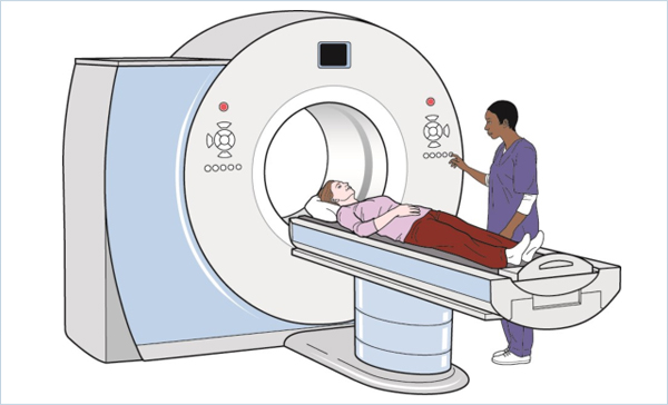
During the procedure
A CT scanner is shaped like a large doughnut standing on its side. You lie on a narrow table with a motor that slides through the center of the scanner into a tunnel. Straps and pillows may be used to help you stay in place. During a head scan, the table may be fitted with a special cradle that holds your head still.
While the table moves you into the scanner, the X-ray tube rotates around you. Each time it rotates, it gives images of thin slices of your body. You may hear buzzing and whirring noises.
A healthcare professional called a CT technologist sits in another room and can see and hear you. You can talk with the technologist through an intercom. To help you keep still during the scan, the technologist might ask you to hold your breath at certain points. Movement can blur the images.During the test, you’ll usually lie on your back on a table (like a bed). If your test requires it, a healthcare provider may inject the contrast dye intravenously (into your vein). This dye can make you feel flushed or give you a metallic taste in your mouth.
When the scan begins:
- The bed slowly moves into the doughnut-shaped scanner. At this point, you’ll need to stay as still as possible because movement can blur the images.
- You may also be asked to hold your breath for a short period of time, usually fewer than 15 to 20 seconds.
- The scanner takes pictures of the area your healthcare provider needs to see. Unlike an MRI scan (magnetic resonance imaging scan), a CT scan is silent.
- When the exam is over, the table moves back out of the scanner.
How long does a CT scan take?
CT scans usually take about an hour. Most of that time is for the preparation. The scan itself takes fewer than 10 or 15 minutes. You can resume normal activities after your provider gives you the OK — usually after they complete the scan and make sure the images are of good quality.
Are there any CT scan side effects?
CT scans themselves usually don’t cause side effects. But some people develop minor side effects from contrast material. These side effects may include:
- Nausea and vomiting.
- Headaches.
- Dizziness.
After the procedure
After the exam you can return to your regular routine. If you were given contrast dye, you may be asked to wait for a short time before leaving to make sure that you feel OK after the exam. You also might be told to drink lots of fluids to help your kidneys remove the dye from your body.
When should I know my CT scan results?
It usually takes about 24 to 48 hours to get the results of your CT scan. A radiologist (a physician who specializes in reading and interpreting CT scans and other radiologic exams) will review your scan and prepare a report that explains the findings. In an emergency setting, like a hospital or emergency room, healthcare providers often receive results within an hour.
Once a radiologist and your healthcare provider have reviewed the results, you’ll either have another appointment or receive a call. Your healthcare provider will discuss the results with you.
Additional Common Questions
Are CT scans safe?
Healthcare providers consider CT scans generally safe. CT scans for children are safe, too. For children, your provider adjusts to a lower dose to reduce their radiation exposure.
Like X-rays, CT scans use a small amount of ionizing radiation to capture images. Possible risks of radiation include:
- Cancer risk: Imaging using radiation, such as X-rays and CT scans, in theory, may cause a slight increase in your risk of developing cancer. The difference is too tiny to measure effectively.
- Allergic reactions: Occasionally, people have an allergic reaction to the contrast agent. This could be a minor or serious reaction.
If you have concerns about the health risks of CT scans, talk to your healthcare provider. They’ll help you make an informed decision about the scan.
Can I have a CT scan if I’m pregnant?
If you’re pregnant or think you might be pregnant, you should tell your provider. CT scans of your pelvis and abdomen can expose the developing fetus to radiation, but it’s not enough to cause harm. CT scans in other parts of your body don’t put the fetus at any risk.
One Final Note..
CT scans are an excellent tool for diagnosing problems with soft tissues, blood vessels, and other body parts that can’t be seen with X-ray or ultrasound imaging. These painless scans don’t require much preparation and can be done quickly in emergency situations. A CT scan takes less than an hour to do, but you may not get results right away, depending on who is interpreting the results. Your doctor will let you know if a contrast dye is necessary for your scan and what action you need to take after the images are evaluated.
It’s normal to have questions or feel a little worried if your provider recommends a CT (computed tomography) scan. But CT scans themselves are painless, carry very little risk and can help providers detect a wide range of health conditions. Getting an accurate diagnosis also helps your healthcare provider determine the best treatment for your situation. Talk to them about any concerns you have, including other options for testing.
Find me on Social Media


 |
Don't forget to bookmark me to see updates.. Copyright © 2000 - 2025 K.
Kerr |